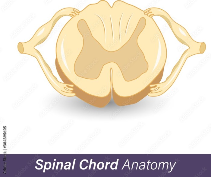
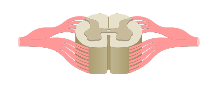
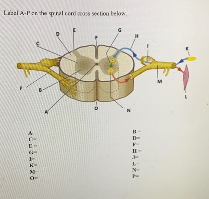
Spinal cord cross section unlabeled – Embarking on a journey into the realm of neuroanatomy, we delve into the intricacies of the spinal cord’s cross-sectional anatomy. This meticulously organized structure serves as the central conduit for communication between the brain and the rest of the body, facilitating both sensory and motor functions.
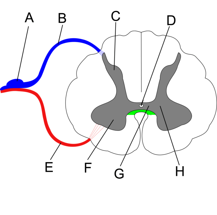
At the heart of the spinal cord lies a distinct division into gray and white matter, each with its specialized roles. The gray matter, shaped like an “H” in cross-section, houses the cell bodies of neurons and is responsible for processing sensory information and initiating motor responses.
In contrast, the white matter, composed primarily of myelinated axons, forms the communication pathways that transmit signals throughout the nervous system.
Cross-sectional Anatomy of the Spinal Cord: Spinal Cord Cross Section Unlabeled

The spinal cord, a cylindrical structure extending from the brainstem to the lumbar region, is composed of gray and white matter. Gray matter, located centrally, forms a butterfly-shaped cross-section. White matter surrounds the gray matter, constituting the outer layer of the spinal cord.The
central canal, a fluid-filled cavity, runs longitudinally through the center of the gray matter. The anterior median fissure, a groove on the ventral surface of the spinal cord, and the posterior median sulcus, a groove on the dorsal surface, further divide the gray matter into left and right halves.
Gray Matter

Gray matter, composed primarily of neuronal cell bodies, dendrites, and unmyelinated axons, is located in the central region of the spinal cord and forms the shape of an “H” or “butterfly.” It is organized into distinct regions that serve specific functions related to sensory and motor processing.
The gray matter is divided into two main regions: the dorsal horns and the ventral horns. The dorsal horns, located on the dorsal (posterior) side of the spinal cord, are responsible for receiving sensory information from the body and transmitting it to the brain.
The ventral horns, located on the ventral (anterior) side of the spinal cord, are responsible for transmitting motor signals from the brain to the muscles.
Substantia Gelatinosa
Within the dorsal horns, there is a specialized region called the substantia gelatinosa. This region is responsible for processing and filtering sensory information, particularly pain and temperature signals. The substantia gelatinosa contains a high concentration of inhibitory neurons that help to modulate the transmission of pain signals to the brain.
White Matter

White matter constitutes the bulk of the spinal cord and is composed of myelinated axons that form ascending and descending tracts. These tracts transmit sensory and motor information to and from the brain.
White matter is organized into three main columns: the dorsal, lateral, and ventral columns. Each column contains ascending and descending tracts that serve specific functions.
Ascending Pathways
Ascending pathways transmit sensory information from the body to the brain. The major ascending tracts in the spinal cord include:
- Posterior column pathway: Transmits fine touch, proprioception, and vibration sense.
- Spinothalamic pathway: Transmits pain, temperature, and crude touch sensations.
- Spinocerebellar pathway: Transmits information about body position and movement to the cerebellum.
Descending Pathways
Descending pathways transmit motor commands from the brain to the spinal cord. The major descending tracts in the spinal cord include:
- Corticospinal pathway: Transmits voluntary motor commands from the cerebral cortex to the muscles.
- Rubrospinal pathway: Transmits postural and movement control signals from the midbrain to the muscles.
- Vestibulospinal pathway: Transmits balance and equilibrium signals from the vestibular system to the muscles.
Meninges and Blood Supply

The spinal cord is enveloped in protective layers of meninges, which consist of three layers: the dura mater, arachnoid mater, and pia mater. The dura mater is the outermost layer and is a tough, fibrous membrane that lines the vertebral canal.
The arachnoid mater is a delicate, web-like membrane that lies deep to the dura mater and contains cerebrospinal fluid (CSF). The pia mater is the innermost layer and is a thin, vascular membrane that closely adheres to the surface of the spinal cord.
The meninges provide several important functions. They protect the spinal cord from mechanical injury, infection, and chemical irritants. They also help to maintain the proper fluid environment around the spinal cord and provide a pathway for blood vessels and nerves to enter and exit the spinal cord.
Blood Supply to the Spinal Cord
The spinal cord receives its blood supply from several arteries, including the anterior spinal artery and the posterior spinal arteries. The anterior spinal artery arises from the vertebral arteries and runs along the ventral surface of the spinal cord. The posterior spinal arteries arise from the vertebral arteries or the posterior inferior cerebellar arteries and run along the dorsal surface of the spinal cord.
The anterior spinal artery supplies blood to the anterior two-thirds of the spinal cord, while the posterior spinal arteries supply blood to the posterior third of the spinal cord. The anterior spinal artery is the main artery supplying the spinal cord, and its occlusion can lead to significant spinal cord damage.
Spinal Cord Reflexes

Spinal cord reflexes are involuntary, rapid responses to stimuli that occur at the level of the spinal cord, without involving the brain. These reflexes are essential for maintaining homeostasis, protecting the body from harm, and coordinating movement.There are several types of spinal cord reflexes, each with a specific function and neural pathway:
Stretch Reflexes
Stretch reflexes, also known as myotatic reflexes, are triggered by the stretching of a muscle. The neural pathway involves a sensory neuron that detects the stretch, an interneuron that relays the signal to a motor neuron, and a motor neuron that stimulates the muscle to contract.
This reflex helps maintain muscle tone and prevents overstretching.
Withdrawal Reflexes, Spinal cord cross section unlabeled
Withdrawal reflexes are triggered by a noxious stimulus, such as pain or heat. The neural pathway involves a sensory neuron that detects the stimulus, an interneuron that relays the signal to a motor neuron, and a motor neuron that stimulates the muscle to withdraw the body part from the stimulus.
This reflex helps protect the body from injury.
Autonomic Reflexes
Autonomic reflexes are involved in regulating involuntary functions such as heart rate, blood pressure, and digestion. These reflexes are mediated by the autonomic nervous system, which consists of the sympathetic and parasympathetic divisions. The sympathetic division is responsible for the “fight or flight” response, while the parasympathetic division is responsible for the “rest and digest” response.
Clinical Significance
Spinal cord reflexes are important for clinical assessment. Abnormal reflexes can indicate damage to the spinal cord or peripheral nerves. For example, an absent stretch reflex may indicate damage to the sensory neuron, interneuron, or motor neuron involved in the reflex arc.
Conversely, an exaggerated reflex may indicate damage to the inhibitory pathways that normally suppress the reflex.
FAQ Summary
What is the significance of the central canal in the spinal cord?
The central canal is a fluid-filled cavity that runs through the center of the spinal cord. It is a remnant of the neural tube from which the spinal cord develops and serves as a conduit for cerebrospinal fluid, which provides nutrients and removes waste products from the central nervous system.
What is the role of the substantia gelatinosa in the spinal cord?
The substantia gelatinosa is a layer of gray matter located in the dorsal horn of the spinal cord. It is responsible for processing and modulating pain signals before they are transmitted to the brain. It plays a crucial role in regulating pain perception and suppressing excessive pain responses.